Radiology imaging stands as a pillar in the medical diagnostic process. With advancements in medical technology, various types of radiology imaging techniques have emerged, each serving a unique purpose in diagnosing and treating disease. This article delves into the various radiology imaging modalities available in modern medicine.
X-ray Imaging
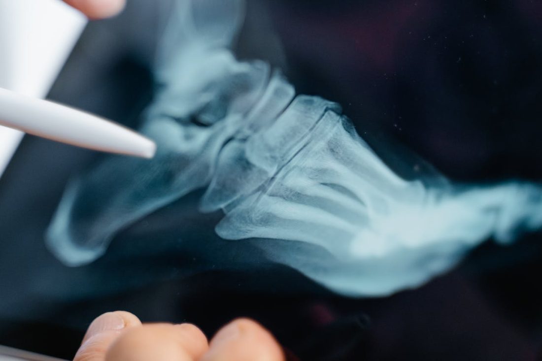
X-ray imaging is a well-known type of radiography. By sending X-ray beams through the body, the varying densities of different tissues can be visualised. Highly dense bones appear white on an X-ray image, softer tissues and fluids appear in shades of grey, and the air in the lungs appears black. This contrast allows for identifying fractures, infections, and abnormal masses.
Computed Tomography
Computed Tomography (CT), also known as “CAT scanning” (Computerised Axial Tomography), provides a more detailed look inside the body than standard X-rays. CT scans use X-rays from multiple angles to create cross-sectional images. These slices can be compiled to form a comprehensive image of areas such as the chest, abdomen, and pelvis, revealing details not visible on a standard X-ray.
Magnetic Resonance Imaging
Magnetic Resonance Imaging utilises strong magnets and radio waves to generate detailed images, particularly of soft tissues. It offers a clear view of body parts that may be obscured on X-rays and CT scans, such as the brain, spinal cord, muscles, and ligaments. MRI is particularly beneficial in neurology, orthopaedics, and oncology for its detailed imaging capabilities.
Ultrasound Imaging
Using sound waves at a high frequency, ultrasound imaging, often called sonography, provides a method to visualise internal body structures. This process involves capturing and transforming echo patterns into visual data, producing live images for analysis. While this technique is prominently used for tracking the progress of pregnancy, it is equally adept at assessing various body organs and tissues, such as the cardiac regions, renal structures, and hepatic tissue.
Nuclear Medicine
Nuclear medicine is a unique imaging modality that assesses function rather than structure. Patients are administered a small amount of radioactive material, which emits radiation as it decays. A special camera detects this radiation and creates images that show how organs function and what they look like. This technique is crucial in cardiology, neurology, and oncology for detecting disease in its early stages.
Positron Emission Tomography
Positron Emission Tomography (PET) scans measure the metabolic activity of cells. Like nuclear medicine studies, PET scans use radioactive substances to create images. By highlighting areas of higher chemical activity, which is often associated with disease processes, PET scans provide valuable information in diagnosing and treating cancer and neurological disorders.
Fluoroscopy
Fluoroscopy is akin to an X-ray “movie.” It is a form of imaging that displays a continuous X-ray image on a monitor, allowing the observation of movement or real-time procedures in the body. This dynamic imaging technique is used in various diagnostic and therapeutic procedures, such as catheter placement and gastrointestinal studies.
Digital Mammography
Digital mammography is an X-ray imaging method specifically designed for breast tissue examination. It is a critical tool in breast cancer detection and uses lower radiation doses than traditional mammography. The images are digitally captured, allowing for enhancements that improve accuracy and detection rates.
The Impact of Diverse Imaging on Patient Care
The diversity of radiology imaging techniques has profoundly impacted patient care. By offering a range of imaging options, radiologists can provide accurate diagnoses and effective treatment planning. For those seeking more information about radiology and its applications, it is worth exploring how each imaging modality caters to different diagnostic needs. Understanding the strengths and limitations of each technique allows healthcare providers to guide patients to the most suitable type of diagnostic imaging, depending on their specific health issues.
Conclusion
The different types of radiology imaging offer a window into the human body, providing invaluable data for medical professionals. This field of medicine has come a long way, from simple X-ray images to intricate scans capable of unveiling comprehensive details about the body’s internal function and anatomy. As technology advances, these imaging techniques continue to evolve, offering even greater potential in diagnosing, treating, and monitoring medical conditions. In tandem with these sophisticated imaging systems, radiology departments rely on specialized equipment like medical grade monitors that meet IEC 60601 certification standards to ensure accurate display and patient‑safe operation.